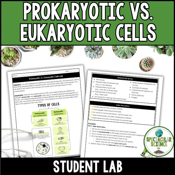
Explore the building blocks of life with this hands-on microscope lab! The Prokaryotic vs. Eukaryotic Cells Lab invites students to investigate cell types by observing real bacterial, plant, and animal cells under a microscope. Students sketch visible structures, classify each type of cell, and dive deep into cell anatomy, structure, and function through guided analysis questions.
Why Teachers Love This Activity:
- Authentic & Engaging Science – Students love the opportunity to use real microscopes and slides, bringing cell theory to life in a tangible and interactive way.
- Perfect for Biology or Life Science – An excellent companion to lessons on cell types, organelles, and the differences between prokaryotes and eukaryotes.
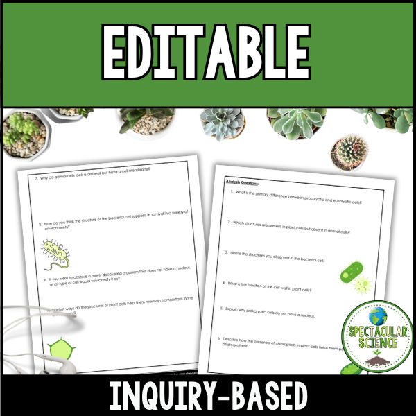
- Encourages Higher-Order Thinking – Students analyze, compare, justify, and imagine—thanks to analysis questions built on all levels of Bloom’s Taxonomy.
- Differentiation-Ready – With labeled vocabulary, observation charts, and drawing opportunities, this lab supports visual, hands-on, and critical-thinking learners.
- Builds Key Lab Skills – Students gain experience using microscopes, identifying cell structures, and carefully documenting scientific observations.
How It Works:
- Set Up & Observe – Students prepare and examine prepared slides of bacterial, plant, and animal cells under a microscope, using appropriate safety and cleaning protocols.
- Draw & Label – Students create labeled sketches of each cell and note key visible structures like the nucleus, cell wall, or flagella.
- Analyze & Compare – Guided analysis questions encourage students to identify cell types, evaluate organelle functions, and compare how different cell structures support life.
- Apply Understanding – Students consider real-life examples, classify unknown organisms, and reflect on how cell structure affects adaptability and energy production.
What’s Included:
- Student Lab Packet with built-in analysis questions
- Answer Key with suggested responses
- Teacher Tips for setup, safety, and guiding student exploration
- Materials List & Safety Guidelines
Why Students Will Love It:
- Microscopes in Action! – Seeing real cells magnified under a microscope makes biology come alive and sparks curiosity.
- Creative Expression – Sketching and labeling what they see helps students connect visual details with scientific terms.
- Feels Like a Real Scientist – Students practice essential lab techniques and get a hands-on feel for how biologists classify and study cells.
- Memorable Comparisons – Distinguishing bacteria, plants, and animals at the cellular level becomes concrete, meaningful, and unforgettable.
Materials (per group):
- Microscope
- Prepared slide of bacterial cells (e.g., E. coli or Bacillus)
- Prepared slide of plant cells (e.g., onion skin or Elodea leaf)
- Prepared slide of animal cells (e.g., cheek cells)
- Lens paper, gloves (optional), goggles
- Water dropper & coverslips (if preparing wet mounts)
- Colored pencils (optional for sketching)
Pro Tip: Introduce the lab with a “cell detective” challenge—give students unlabeled images of cells and have them guess if they’re looking at a prokaryote or eukaryote before starting the lab. This sparks curiosity, encourages close observation, and builds excitement for using the microscope to confirm their hypotheses!
Grade Levels: 8th – 12th
Total Pages: 21
Teaching Duration: 60 Minutes
If you have any questions, please do not hesitate to ask.
Thanks for visiting!
Jessica